Tuesday, October 29, 2013

Lab Observations 3

Made: 10.29.13
       I made the third set of observations on my MicroAquarium today. At first, I tried the compound microscope, but neither Dr. McFarland nor myself could focus the lens correctly. This proved to be the theme of day, unfortunately.
       It turns out that a great many changes had taken place since last week. Yesterday, a beta food pellet was added to the aquarium. When I viewed my aquarium (after the second microscope..) I noticed that quite a lot of activity was happening around said pellet, which had mostly dissolved leaved debris floating throughout the aquarium. This included activity from several different new organisms and new types of organisms. I noted the addition of these new species: Euchlanis sp., Lecane sp. (Pennack, 1953), Vorticella sp., Coleps sp., Cyclops sp.(Rainis & Russell, 1996), Tachysoma sp, Halteria sp., Cercomnas sp., and Anisonema sp. (Patterson, 2003). I also saw a few organisms that were not properly identified, including a worm-like organism that appeared to have a light receptor (like the cyclops) and what I believe is a younger Philodina sp. Some of the organisms were more noteworthy than others. I will describe the behaviors and observations I noted while viewing each organism, respectively, in the following paragraphs.
         I could see movement in my aquarium with the naked eye. This appeared to be the Cyclops sp., of whom I viewed at least three of various sizes, now currently residing in my aquarium (Pennack, 1953). They acted as if the light from the microscope irritated them, so they swam away as the slide moved over it. This made watching them a tad difficult though. A few different specimens of the Euchlanis sp. and the Lecane sp. were captured and observed together. The movement of the Euchlanis sp. as well as its appearance reminded me of the Philodina sp., which makes sense as they are both rotifers.(Pennack 1953).They (as well as most of the organisms) tended to dwell near the food pellet and surrounding debris. However, two larger Euchlanis sp.'s were viewed swimming along the bottom of the tank as well, one of which seemed to eat a smaller organism, but it could have been debris. Speaking of the bottom of the tank, I did not observe any dead organisms, but I'm sure there must be some by now. I didn't see the Philodina sp. from last time, so it may very well be dead, just unseen (Pennack 1953).
      There were many Coleps sp. in my aquarium today. They're just small, black, spherical organisms, one of which appeared to be ingesting particles/debris from its surroundings (Rainis & Russell 1996).  I managed to catch pictures of the Tachysoma sp. (which had a spastic movement), the Halteria sp. (Patterson 1996), and, my favorite of today, the Vorticella sp, (Rainis & Russell 1996). These organisms are documented in Fig 1, 2, and 3, respectively.

Fig. 2. Shows Halteria sp. notice cilia
 protruding all around, like a starburst.
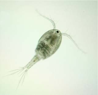
Fig. 1. Shows Tachysoma sp. note "feelers"
protruding from tail end (right).


Fig. 3. The Vorticella sp. note long protrusion attached
to plant and bell like body (Rainis & Rusell 1996).
         The Vorticella sp. was particularly interesting. I learned they usually are found in clusters or groups (Rainis & Russell 1996). I know I personally saw around four in the same area (is that a "cluster"?). I found their bell shape appealing and the way they moved resembled seaweed, as in they swayed. Very cool organisms.
        There are both single and multicellular organisms in my micro-aquarium, though as we've seen the organism types vary each time. Each of these organisms move, but their movements also vary, ranging from quick bursts to slow spins. There are chlorophyll organisms in the form of plants, though the plants aren't doing so well in this environment. They look basically dead. They've aged and turned dark. Algae-like debris has started to collect on the prongs of the Ultricularia gibba plant (McFarland, 2013). This could, however, be particles of the almost fully dissolved betta food pellet.

Tuesday, October 22, 2013

Lab Observations 2

Made: 10.22.13
     My second set of MicroAquarium observations were made today, first using the light microscope and later, the camera/microscope. Immediate noticeable changes in the MicroAquarium were made. Utricularia gibba, the plant, has now taken on a slightly browned appearance (McFarland, 2013).  The aquarium also went through several changes with the "non-green" organisms. First of all, no organisms were observed that had died and sank to the bottom (luckily), but new organisms have appeared. This includes the addition of at least one Philodina sp., a single celled organism I will discuss momentarily (Pennack,1953). There may have been even another type of rotifer which I was unable to capture to identify properly so the  identification may be incorrect. Also, I should note that my lab instructor pointed out a possible "Carpenter's Ruler", a thin stick-like organism, however no movement was observed and a proper identification was not mad to prevent the possibility of incorrect identification. These new organisms tended to the bottom of the aquarium, where the debris lay.  The single cyclops, now assuredly  identified from the previous observations taken, has gained a faster and more sporadic pattern of movement, so much so I was almost unable to view it (Rainis & Russell,1996). In the case of the many flagellates/ciliates (first sited 10/17), while the speed of the organism stayed relatively the same, they had a more varied tendency of location.
      The Philodina sp., which I referred to earlier, not only had a very strange movement, which consisted of what appeared to be eating the dirt/debris extremely rapidly, but was also hard to identify and observe. When I first spied what I BELIEVE to be this organism, I showed it to the instructor who identified it as a rotifer. A bit later, I noticed it again. It had moved locations without leaving the debris area ( I could have mistaken two completely separate organisms). However, it appeared larger and was "eating" more rapidly than originally.  The "stomach" area and in fact, the whole organism was rather transparent which allowed me to see literally inside the organism as it ingested the debris (Fig. 1). The appearance of it was interesting, indeed. The lab instructor and I looked up a diagram of Philodina sp., and compared it to the one in my MicroAquarium. We noticed that mine seemed to have three or four "toes" while the average specimen has two (Fig 2.). This, accompanied with the large size and strange movements, led me to hypothesize that the organism was currently participating in asexual reproduction. After a bit of background research on Philodina sp., I found that, "Several types of reproduction have been observed in rotifers. Some species consist only of females that produce their daughters from unfertilized eggs, a type of reproduction called parthenogenesis. In other words, these parthenogenic species can develop from an unfertilized egg, asexually (Speer, 2000)." This leads me to further believe that it is likely the specimen was exhibiting the above  process and perhaps new specimen of Philodina sp. will be available in my aquarium for observing next week.
              I managed to capture a couple of pictures of the Philodina sp. using the cameras attached to the light microscope. The pictures are documented below as Fig 1. and Fig. 2. Notice the small tentacle-like protrusions in Fig. 1. These were in constant motion- ingesting debris or not.
Fig. 1. Shows Philodina sp. with ingested debris and
tentacle-like protrusions on left near mouth (Pennack, 1953).


Fig. 2. Shows Philodina sp. specimen with multiple "toes" (Pennack, 1953).

Thursday, October 17, 2013

Initial Set-Up

Made: 10.17.13
         The initial set-ups of our MicroAquariums were yesterday. In the lab room, each of us were seated at lab benches with samples of water and "debris" from various river locations around Knoxville; attached to which was a pipet. Also on the lab bench was a light microscope. We were each given the pieces to a MicroAquarium which included a lid, stand holder, and small glass tank. On the face of the small glass tank we were instructed to label our MicroAquariums using small colored stickers. After our MicroAquariums were properly labeled, we used a pipet to extract water from the containers on the lab benches. I chose water sample 8 or, water gathered from the Tennessee River at the boat ramp across from the Knoxville sewer plant (Neyland Dr. Knox Co. Knoxville TN. Full sun exposure. French Broad and Holston Rivers water Sheds N35 56.722 W83 55.587 813 ft 10/13/2013) (McFarland, 2013). First, water and debris from the bottom of water sample 8 was obtained with the pipet and transferred to the MicroAquarium. Next, water from the sample coming from the middle was added, and finally water was taken from the top and added to the MicroAquarium (Cook & McFarland, 2013).
            Samples of Amblestegium varium (Hedwig) Lindberg (Moss. Collection from: Natural spring. at Carters Mill Park, Carter Mill Road, Knox Co. TN. Partial shade exposure. N36 01.168 W83 42.832. 10/13/2013), Fontinalis sp. (Moss.  Collected from: Holston River along John Sevier Hwy under I 40 Bridge Partial shade exposure Holston River water Shed N36 00.527 W83 49.549 823 ft 10/13/2013) and Utricularia gibba (L. Flowering plant. A carnivous plant. Original material from south shore of Spain Lake (N 35o55 12.35" W088o20' 47.00), Camp Bella Air Rd. East of Sparta Tn. in White Co. and grown in water tanks outside of greenhouse at Hesler Biology Building. The University of Tennessee. Knox Co. Knoxville TN. 10/13/2013) were available for adding to our MicroAquariums (McFarland, 2013). I added a small amount of each to my tank. Our lab instructor enlightened the class and revealed that the mosses were added to oxygenate the water, which any organisms in our tank will enjoy.
Fig 1. Shows the micro-invertebrate,
 "Cyclops", the same type of organism seen
through my microscope (Schur, 2009)
           After the MicroAquariums were fully assembled, the first observations could be made using a light microscope. Observations made using the 10X scope worked best for me. I observed several moving organisms which appeared to be the freshwater protistas, flagellates and ciliates (single-celled), and just one of the micro-invertebrate- "cyclops". These identifications are rough and improper. The flagellates and ciliates appeared to be moving in a haphazard circular fashion, while the cyclops had a quick movement that was more controlled. The cyclops at one point appeared to be eating or digesting particles of the debris within the tank. The cyclops tended to dwell near the bottom of the tank with the debris, while the flagellates/ciliates dwelled nearer to the added mosses (middle of tank).
           I did not personally take any photographs of the organisms in my MicroAquarium, but I obtained an image from the web of a cyclops to show as a reference. The example is pictured above labeled Fig. 1.

Bibliography

Branson, David. "Zooplankton of the Great Lakes" [Internet] Central Michigan University; c2009              [cited 2013 Nov 11]. Availible from:                                                                                         http://people.cst.cmich.edu/mcnau1as/zooplankton/20web/Craspedacusta2/Craspedacusta.htm
Cook R. and McFarland K. 2013. General Botany 111 Laboratory Manual.15th edition.

          University of Tennessee. p. 155-156 
McFarland, Kenneth [Internet] Botany 111 Fall 2013. [cited 10/16/2013]. Available from:                         http://botany1112013.blogspot.com/
Patterson, David J. 1996. Free-living freshwater protozoa: a colour guide. New York: Wiley. p. 
54,
           65, 95, 125, 175, 305

 Pennack, Robert W. 1953. Fresh-water Invertebrates of the United States. New York: Ronald                   Press Company. p.  162, 170, 197                                                                      
Pennack, Robert W. 1989. Fresh-water Invertebrates of the United States. 3rd Edition. 
           New York: Ronald Press Company. p. 116 
Rainis KG and Russell BJ. 1996.  Guide to Microlife. Danbury Connecticut: Grolier Publishing. p.
           85, 105, 109, 203, 217
Schur, Chris & Dawn. "One-eyed Monsters" [Internet] Schur's Astro Photography;
           c2009. 
[modified 2009 Apr 2; cited 2013 Oct. 16]. Available from:
           
<http://www.schursastrophotography.com/robotics/visionlogic-4.html>
Speer, Brian R., et al. "Introduction to the Rotifera". [Internet]. Berkeley University; c2000.
            [modified 2000 Jun. 8; cited 2013 Oct. 22]. Available from:
            <http://www.ucmp.berkeley.edu/phyla/rotifera/rotifera.html>