The initial set-ups of our MicroAquariums were yesterday. In the lab room, each of us were seated at lab benches with samples of water and "debris" from various river locations around Knoxville; attached to which was a pipet. Also on the lab bench was a light microscope. We were each given the pieces to a MicroAquarium which included a lid, stand holder, and small glass tank. On the face of the small glass tank we were instructed to label our MicroAquariums using small colored stickers. After our MicroAquariums were properly labeled, we used a pipet to extract water from the containers on the lab benches. I chose water sample 8 or, water gathered from the Tennessee River at the boat ramp across from the Knoxville sewer plant (Neyland Dr. Knox Co. Knoxville TN. Full sun exposure. French Broad and Holston Rivers water Sheds N35 56.722 W83 55.587 813 ft 10/13/2013) (McFarland, 2013). First, water and debris from the bottom of water sample 8 was obtained with the pipet and transferred to the MicroAquarium. Next, water from the sample coming from the middle was added, and finally water was taken from the top and added to the MicroAquarium (Cook & McFarland, 2013).
Samples of Amblestegium varium (Hedwig) Lindberg (Moss. Collection from: Natural spring. at Carters Mill Park, Carter Mill Road, Knox Co. TN. Partial shade exposure. N36 01.168 W83 42.832. 10/13/2013), Fontinalis sp. (Moss. Collected from: Holston River along John Sevier Hwy under I 40 Bridge Partial shade exposure Holston River water Shed N36 00.527 W83 49.549 823 ft 10/13/2013) and Utricularia gibba (L. Flowering plant. A carnivous plant. Original material from south shore of Spain Lake (N 35o55 12.35" W088o20' 47.00), Camp Bella Air Rd. East of Sparta Tn. in White Co. and grown in water tanks outside of greenhouse at Hesler Biology Building. The University of Tennessee. Knox Co. Knoxville TN. 10/13/2013) were available for adding to our MicroAquariums (McFarland, 2013). I added a small amount of each to my tank. Our lab instructor enlightened the class and revealed that the mosses were added to oxygenate the water, which any organisms in our tank will enjoy.
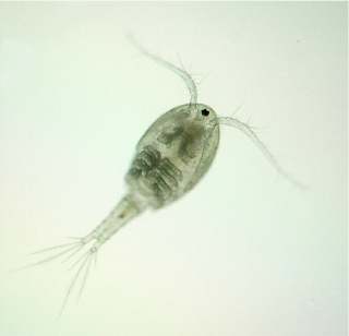 |
| Fig 1. Shows the micro-invertebrate, "Cyclops", the same type of organism seen through my microscope (Schur, 2009) |
I did not personally take any photographs of the organisms in my MicroAquarium, but I obtained an image from the web of a cyclops to show as a reference. The example is pictured above labeled Fig. 1.
No comments:
Post a Comment